East Melbourne VIC 3002

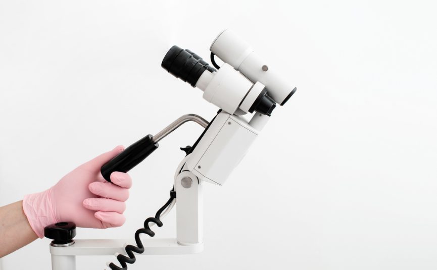
Understanding changes or symptoms affecting the vulva can be difficult, especially when many conditions are not visible to the naked eye. That’s where vulvoscopy comes in — a specialised diagnostic procedure designed to examine the vulval skin in exceptional detail. Much like a colposcopy used to assess the cervix, vulvoscopy uses a magnifying instrument called a vulvoscope to closely inspect the external genital area.
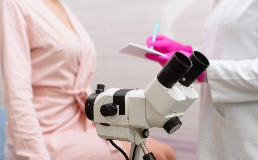
What is a Vulvoscope?
A vulvoscope is an operating microscope which allows the visualisation of the vulval skin and surrounding structures in great detail. It magnifies the area and projects it onto a screen that you the patient, can see. This allows us to point out any abnormalities if you want to see what is happening, some patients prefer not to and in that case, the screen is turned away.
Besides magnifying the skin for any abnormalities, there are a number of filters which allow us to better visualise structures such as blood vessels, which may be important in a diagnosis. Magnification via a vulvoscope allows lesions that are not visible with the naked eye to be seen and defined.
When is Vulvoscopy Recommended?
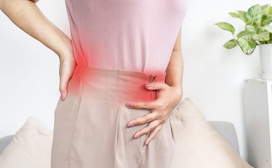
The vulvoscopy procedure is performed in any patient who has a suspicion of a vulval skin disorder, a vulval symptom such as itching, burning or pain or a lesion on the vulva which needs a diagnosis.
The vulvoscopy instrument is a modern digital microscope and it is built into a comfortable chair which supports the patient. The patient has a gown on, is covered by a blanket and there is a registered nurse present to chaperone and help the patient and to provide any assistance for A/Prof Kliman that are required such as swabs or biopsies.
The procedure itself is totally painless. Occasionally a vulval biopsy will be required and this is a punch biopsy with a diameter of 1 mm. It is performed after a strong local anaesthetic is injected with a very fine 30 gauge needle into the skin to make the area completely numb. Associate Professor Kliman forwards all his biopsies to Professor James Scurry at the University of New South Wales who is a world expert when it comes to vulval skin pathology.
Benefits of Vulvoscopy Procedure
The advantages of using a vulvoscope is that it allows for a more accurate diagnosis, for skin lesions to be more accurately assessed and it allows for careful decision making with respect to where is the best location to take a biopsy.
Any precancerous lesion which may occur with certain skin diseases, can be identified when it is particularly small and readily treatable.
Vulval biopsies are taken where skin changes could be due to a number of possible conditions or where a lesion or skin disease has features suggesting precancerous change.
If you require a biopsy, the area will remain numb for some time, the very small biopsy site is covered with an antiseptic and you will be given simple instructions on how to care for that area of your skin.
It is important to realise that at all times, your privacy and dignity are respected as one would expect.
A/Prof Kliman’s Diagnostic Approach Using a Vulvoscope
Basically, the use of a vulvoscope allows Associate Professor Kliman to better define and diagnose conditions such as lichen sclerosus, lichen planus, psoriasis and lesions such as ulcers or pigmented lesions. That means the precision involved with defining a vulval skin disorder or a vulval lesion is substantially increased, especially in patients who have longstanding disorders or symptoms such as itch, burning, irritation or vulval pain.
Read More: Patient Recommendations After Vulvar Biopsy